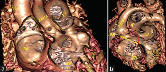
Figure 5.

Virtual cardiac dissection from a 20-month-old girl with double outlet right ventricle shows the morphology of the subaortic interventricular communication (red dotted line) in anteroposterior (a) and left anterior oblique projection (b). (a) In the anteroposterior plane, it shows the defect opens to the right ventricle directly beneath the aortic root. (b) In the left anterior oblique projections, it shows that the area (yellow double-headed arrow) around which the surgeon will create a tunnel to reconnect the aortic root with the left ventricle is analogous to the ventricular septal defect as seen in the setting of overriding of the aortic root [Figure 4]. In reality, the area currently described as the “ventricular septal defect” is the outlet for the morphologically left ventricle (red double-headed arrow). Virtual dissection shows why it is more appropriate to use the term interventricular communication in the setting of double outlet right ventricle. LA: Left atrium, LV: Left ventricle, RA: Right atrium, RV: Right ventricle, PT: Pulmonary trunk, SCV: Superior caval vein
