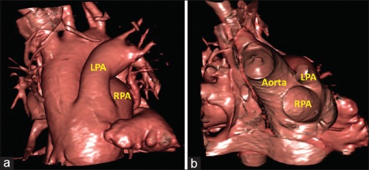
Figure 6.

Virtual cardiac dissection obtained from a patient with common arterial trunk. It is not possible from the external view (a) to demonstrate the precise arrangement of the orifices of the left and right pulmonary arteries. The internal view (b), in anteroposterior projection with cranial tilt, on the other hand, provides an unequivocal demonstration of the closely separated locations of the orifices of the pulmonary arteries. LPA: Left pulmonary artery, RPA: Right pulmonary artery
