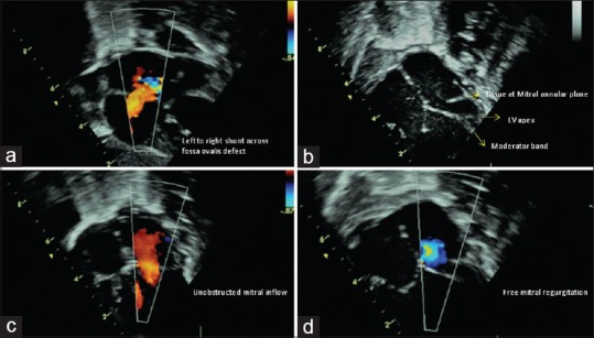
Figure 1.

(a) Subcostal view showing the left-to-right shunt across fossa ovalis defect. (b) Apical four-chamber view in diastole. Arrow points to the lateral ridge of tissue at the plane of the mitral annulus. No valve is seen. Dashed-arrow points to the intact ventricular septum and shows the left ventricle reaching half way to the apex formed by the right ventricle. Bold arrow shows the moderator band in the right ventricle with its septal and parietal attachments. (c) Unobstructed laminar mitral inflow. (d) Free mitral regurgitation
