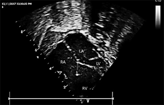
Figure 2.

An enlarged view showing the crux of the heart with intact ventricular septum, a small left ventricle, and ridge of tissue on the lateral aspect of the mitral annular plane. Right atrium, left atrium, dilated right ventricle, and hypoplastic left ventricle are labeled within the figure
