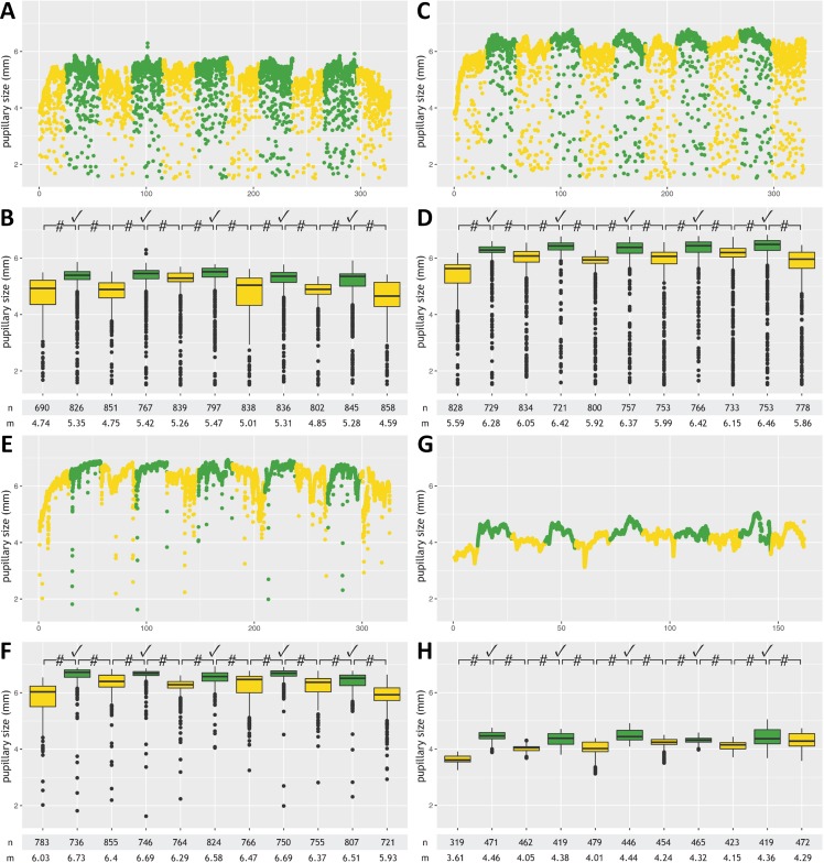
Figure 2. Pupillometry data from four patients with successful command following.
This figure shows results from participants with successful command following, detected by automated pupillometry, during a mental arithmetic paradigm: two patients admitted to the neurological ward with diagnoses of multifocal motor neuropathy (A, B) and Guillain-Barré syndrome (C, D), respectively, a healthy participant (E, F), and a conscious 34-year old male with the pharyngeal-cervical-brachial variant of Guillain-Barré syndrome admitted to the ICU (G, H). Data from each subject are presented twice and in 2 different formats (raw measurements A, C, E and G; annotated data B, D, F, H). Minor artifacts due to blinking or eye movements are seen in A, C and E, but not G (probably because of facial and oculomotor nerve palsies). Color code: Periods with mental arithmetic are shown in green, rest periods in yellow. Numbers on the x-axis (“0-100-200-300”) denote time in seconds. Pupillary sizes during mental arithmetic were significantly larger (p-value <0.0001) than during rest periods, consistent with pupillary dilation, in all five tasks for each of the four participants. #, p-value <0.0001; ✓, pupillary dilation; n, number of measurements; m, median pupillary size; mm, millimeter.

