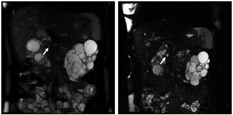
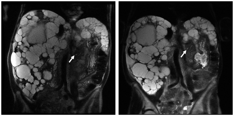
FIGURE 2.
Pancreatic Cyst Lesions (PCLs) From Magnetic Resonance Imaging (MRI) of Patient With PKD1 and PKD2 Genes. A, Coronal single-shot fast-spin echocardiography (SSFSE) MRI showing a PCL (arrow) in 28-year-old woman with PKD1 from 2006. B, Coronal SSFSE MRI showing increased PCL size (arrow) in the same woman with PKD1 in 2011. C, Coronal SSFSE and coronal half-Fourier acquired single-shot turbo-spin echocardiography (HASTE) MRI showing a PCL (arrow) in the tail of the pancreas of a 65-year-old woman with PKD2 from 2006. D, Coronal SSFSE and coronal HASTE MRI showing increase in PCL size (arrow) from 2011.


