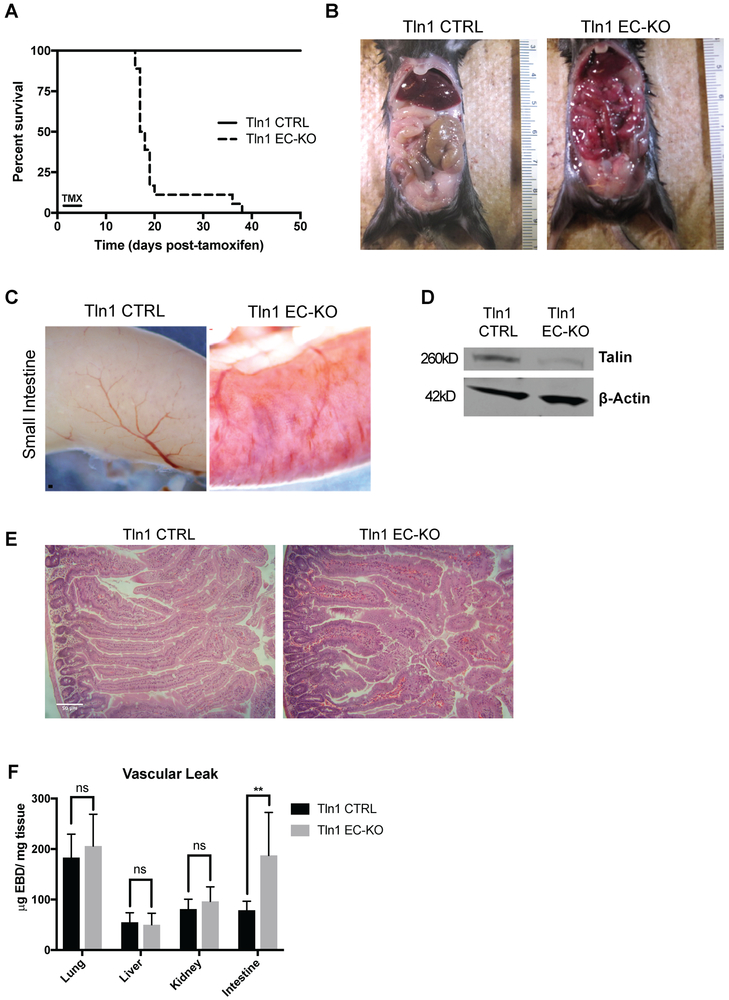
Figure 1: Endothelial cell-specific deletion of talin1 in established blood vessels causes intestinal vascular hemorrhage and death.
A-C. Adult Tln1 EC-KO (Tln1fl/fl;Cdh5creERT2+/−) and Tln1 CTRL (Tln1fl/fl;Cdh5creERT2−/−) mice were administered tamoxifen once a day for 3 consecutive days via intraperitoneal injection. A. Survival of Tln1 EC-KO and Tln1 CTRL mice following tamoxifen treatment (n=15, Tln1 CTRL; n=14, Tln1 EC-KO). B. Pictures of exposed peritoneum of adult Tln1 EC-KO and CTRL mice sacrificed 16 days after tamoxifen treatment. C. Macroscopic images of intestinal vascular hemorrhage in Tln1 EC-KO adult mice 16 days after tamoxifen treatment. D. Western blot analysis of talin expression in mouse lung endothelial cell cultures isolated from Tln1 CTRL or Tln1 EC-KO mice. Cultures were treated with 4-hydroxy-tamoxifen for 4 days and protein lysates subjected to Western blotting with talin and β-actin antibodies. (n=2). E. Hematoxylin/eosin staining of small intestine isolated from Tln1 CTRL and Tln1 EC-KO mice 16 days after tamoxifen treatment showing the presence of extravascular red blood cells in Tln1 EC-KO villi. (n=3; scale=50 μm). F. Measurements of Evan’s Blue Dye (EBD) in lung, liver, intestine, brain and kidney of Tln1 CTRL and Tln1 EC-KO mice 2 hours after intravenous injection. (n=12, Tln1 CTRL; n=10, Tln1 EC-KO; **p = 0.0013 two-tailed unpaired t-test).

