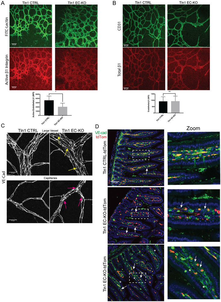
Figure 3: Reduced β1 integrin activation and disorganized adherens junctions in established vessels of Talin1 EC-KO mice.
A-B. Immunofluorescence analysis of active β1 integrin with the activation-sensitive antibody 9EG7 (A) or total β1 integrin with an activation insensitive β1 integrin antibody HMb1-1 (n=3-4; *p=0.03 two-tailed unpaired t-test) (B) in whole mounted retinas from Tln1 EC-KO and Tln1 CTRL mice. Neonates were treated with tamoxifen on P1-3 and sacrificed on postnatal day 7 (P7) at which time retina whole mounts were prepared for staining. (n=3-4; ns=not significant unpaired t-test; scale=50 μm). C. Altered junctional thickness (magenta arrows) and localization of VE-Cadherin (yellow arrows) in retinal vessels and capillaries of Tln1 EC-KO mice 16 days after tamoxifen injections compared to Tln1 CTRL mice. (n=3; scale=25 μm). D. VE-cadherin immunofluorescence of intestine cryosections showing disrupted cell-cell junctions in villi (white arrows) of Tln1 EC-KO-tdTom mice 16 days after tamoxifen treatment. Changes in cell-cell junctions appear cell autonomous as junctions between non-recombined ECs in Tln1 EC-KO-tdTom villi (asterisks) are intact. (n=3; scale=100 μm).

