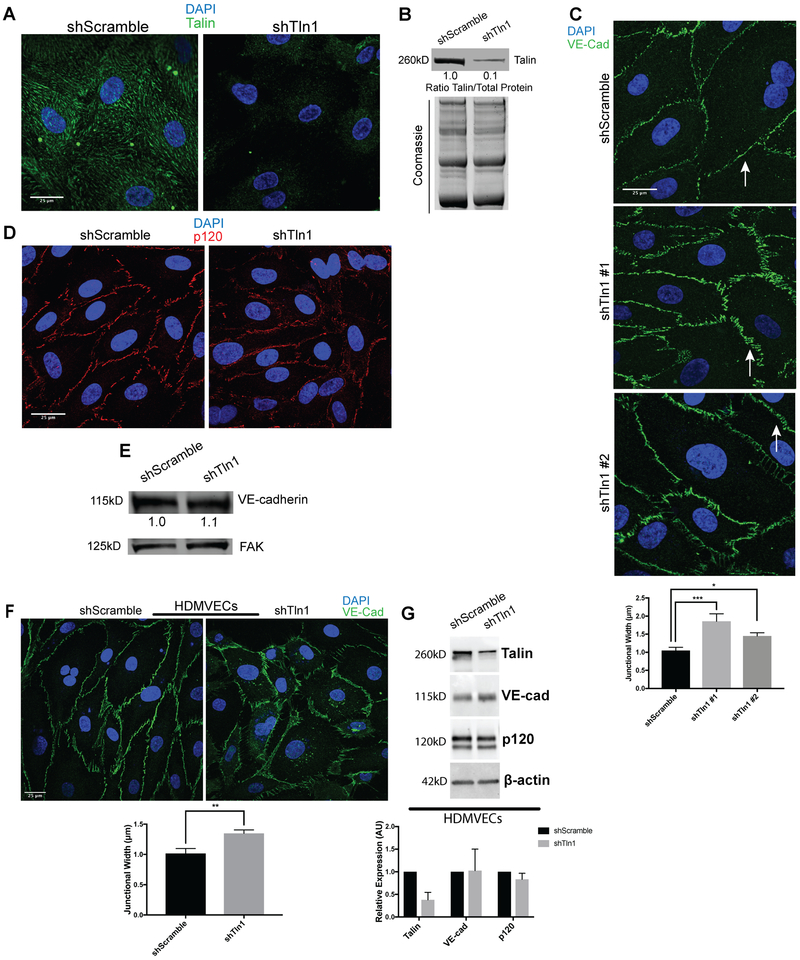
Figure 4: Increased width of adherens junctions formed by talin-deficient endothelial cells.
A. HUVECs were infected with talin1 shRNA (shTln1) or scramble sequence shRNA (shScramble) lentivirus. Immunostaining for talin1/2 72 hours after shRNA infection shows efficient knockdown of talin protein in shTln1 cells (n=5; scale=25 μm). B. Western blot analysis and quantitation of talin1 protein levels relative to total protein in shScramble and shTln1 treated HUVECs (n=5). C. Max-intensity projections depicting Immunofluorescence of VE-Cadherin in cells infected with two different shTln1 lentiviruses. shTln1 HUVECs shows disorganized cell-cell junctions and increased junctional width (white arrows) compared to shScramble HUVECs. (n=3; ***p=0.0007, *p=0.0225 one-way ANOVA with Dunnett’s multiple comparisons test). D. Immunofluorescence of p120-catenin in shTln1 and shScramble HUVECs. (n=3). E. Western blot analysis of VE-cadherin protein levels in shScramble and shTln1 HUVECs show similar levels of expression (n=6). F. Human dermal microvascular cells (HDMVECs) treated with shTln1 lentivirus exhibit increased junctional width relative to shScramble treated cells (n=3; scale=25 μm; **p=0.0042 two-tailed unpaired t-test). G. Western blot analysis and quantitation of talin1/VE-cad/p120 protein levels in shScramble and shTln1 HDMVECs. Efficient deletion of talin1 does not appear to affect relative expression of VE-cad or p120 protein (n=3).

