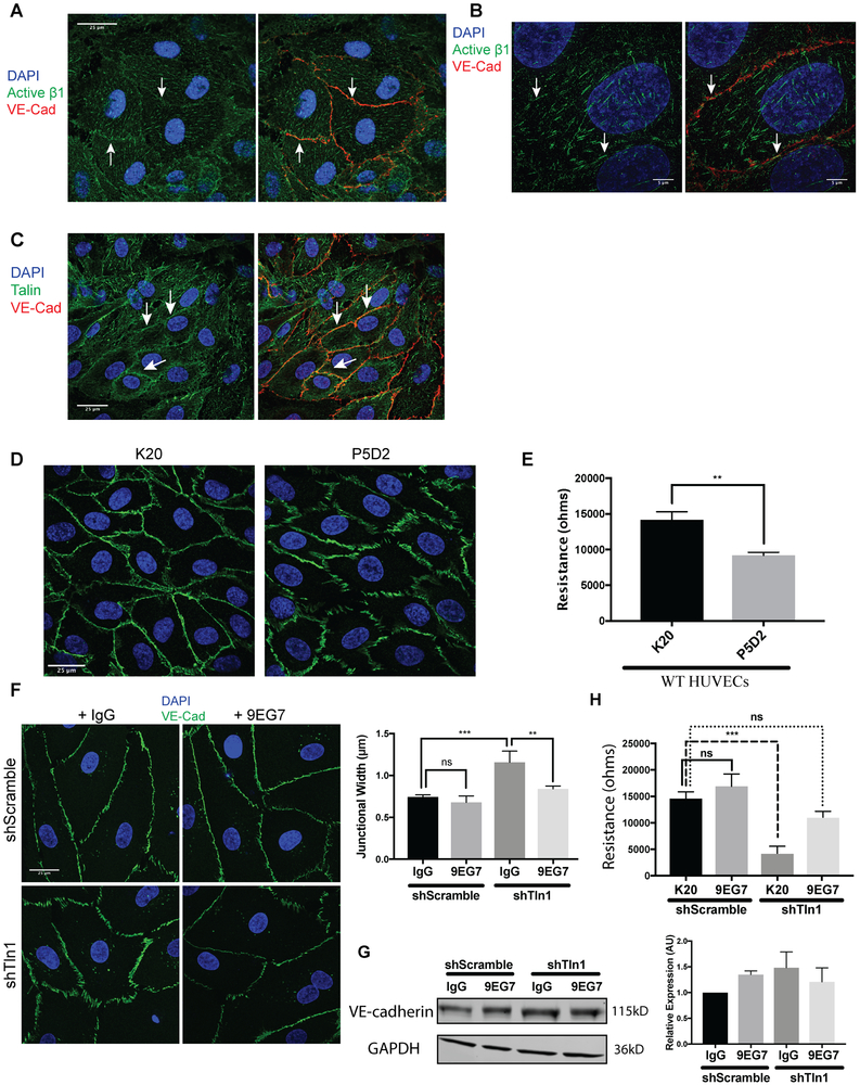
Figure 6: Talin-dependent β1-integrin activation is required for endothelial barrier function.
A. Z-stack projections of immunofluorescence using an activation-sensitive β1 integrin antibody (9EG7) (green) and VE-Cadherin antibody (red) on HUVECs showing the appearance of a subpopulation of β1 integrin near AJs of confluent HUVECs. White arrows highlight cell-cell border regions enriched for β1 integrin. (n=3; scale=25 μm). B. Super resolution 3D structured illumination (3D-SIM) immunostaining of active β1 integrin and VE-cadherin in HUVECs highlight a subset of β1 integrin (white arrows) at VE-cadherin-positive junctions (n=3; scale=5 μm). C. Immunofluorescence on HUVECs with antibodies against talin (green) and VE-cadherin (red). A subset of talin localized to cell-cell contacts (white arrows) in addition to the expected pool of talin at focal adhesions (n=3; scale=25 μm). D. Treatment of HUVEC monolayers with a β1 integrin blocking antibody (P5D2) alters cell-cell junction organization relative to cells treated with a non-function altering β1 integrin antibody (K20). (n=3; scale=25 μm). E. HUVEC monolayers treated with a β1 integrin blocking antibody (P5D2) exhibit reduced barrier function as measured by electrical cell-substrate impedance sensing (4000 Hz) relative to HUVECs treated with a non-function altering β1 integrin antibody (K20). Measurements were made 3 hours after antibody incubation and remained stable for up to 6 hours post-treatment. (n=3; p=0.0019 unpaired t-test). F. Junctional width measured by VE-Cadherin immunofluorescence (green) is normalized by antibody-mediated β1 integrin activation (9EG7) in shTln1 HUVECs relative to talin-deficient HUVECs treated with a non-function altering antibody (K20). (n=3; scale=25 μm; *** p=0.0006, ** p=0.0036 ordinary one-way ANOVA with Sidak’s multiple comparisons test). G. VE-cadherin protein expression measured by western blot in shScramble and shTln1 HUVECs treated with 5 μg/mL Rat Isotype IgG or β1 integrin activating antibody 9EG7 for 12 hours. (n=2; ns; One-way anova with a Tukey multiple comparisons test) H. Electrical resistance of shScramble and shTln1 HUVECs treated with either the activating β1 integrin antibody 9EG7 or the non-function altering β1 antibody K20. (n=3; *** p=0.0002 one-way ANOVA with a Tukey multiple comparisons test).

