Figure.
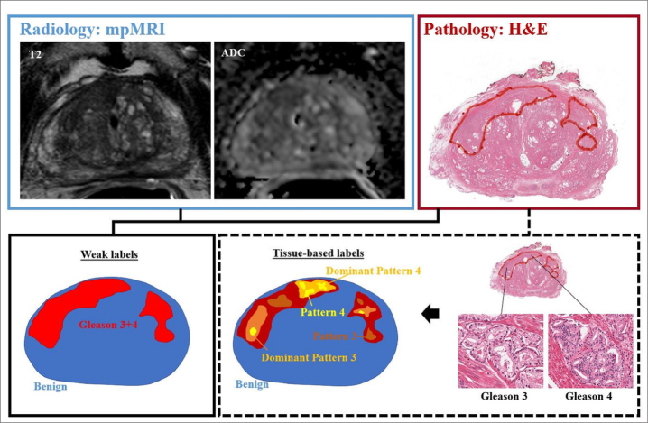
Example mpMRI (T2-weighted and ADC images) with corresponding whole mount hematoxylin-eosin (H&E) section demonstrating two pathologically-defined tumors. Overall assessment in this patient resulted in Gleason grade 3+4 score assignment, resulting in weak labels derived from total tumor extent. However, detailed pathologic assessment demonstrates majority of Gleason pattern 4 is located in the anterior portion of the tumor, with predominately Gleason pattern 3 throughout remainder of the tumor field. Opportunities for improved spatial learning include density mapping of dominant pathologic grading and exclusion of non-cancerous structures within tumor field.

