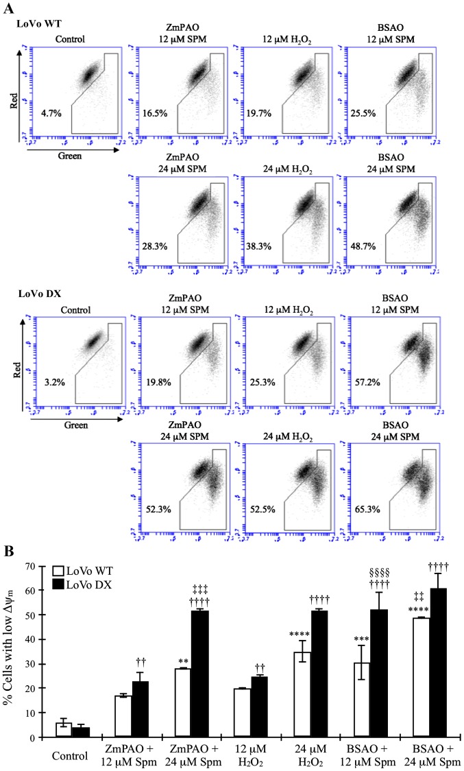
Figure 10.
Effects of the treatment with ZmPAO or BSAO and spermine on mitochondrial membrane potential in LoVo cells. LoVo WT and LoVo DX cells were treated with various doses of hydrogen peroxide (positive control) or spermine in the presence of ZmPAO or BSAO for 1 h. JC-1 fluorescent staining intensity was examined using flow cytometry. (A) Results are shown as dot plots and as the percentages of cells with an energized (top) or depolarized (bottom) mitochondrial membrane. Data are representative of 2 independent experiments with similar results. (B) Each bar represents the mean ± SD of the cells with depolarized mitochondria of 2 independent experiments. Data were analyzed by one-way ANOVA, followed by Tukey’s post hoc test. **P<0.01, ***P<0.001 and ****P<0.0001 vs. control LoVo WT cells; ††P<0.01 and ††††P<0.0001 vs. control LoVo DX cells; ‡‡P<0.01 and ‡‡‡P<0.001 vs. LoVo WT cells incubated with ZmPAO and 24 µM spermine; §§§§P<0.0001 vs. LoVo DX cells incubated with ZmPAO and 12 µM spermine. ZmPAO, maize polyamine oxidase; BSAO, bovine serum amine oxidase; LoVo WT cells, LoVo wild-type cells; LoVo DX cells, LoVo multidrug-resistant cells.

