Figure 6.
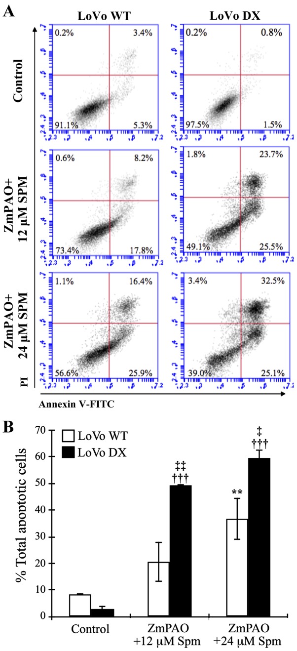
Flow cytometric analysis of apoptosis of LoVo cells after double labeling with Annexin V-FITC and PI. LoVo cells were incubated with 12 or 24 µM spermine in the presence of ZmPAO for 60 min at 37°C. At 24 h after the end of the treatment, and incubation at 37°C, cells were analyzed by flow cytometry. (A) Representative Annexin V-FITC and PI flow cytometry dot plots of LoVo WT and LoVo DX are shown. The x-axis represents FITC staining, and the y-axis represents PI staining. The percentage of cells displaying Annexin V-FITC positive/PI-negative (early apoptosis), Annexin V-FITC positive/PI-positive (late apoptosis or dead), Annexin V-FITC negative/PI-positive (necrosis) and double negative cells (viable cells) is indicated. The dot plots have been obtained from 1 out of 2 independent experiments, performed in the same experimental conditions, which gave similar results. (B) Each bar represents the mean ± SD of total apoptotic cells of 2 independent experiments. Data were analyzed by one-way ANOVA, followed by Tukey’s post hoc test. **P<0.01 vs. control LoVo WT cells; †††P<0.001 vs. control LoVo DX cells; ‡P<0.05 and ‡‡P<0.01 vs. LoVo WT cells incubated with ZmPAO and spermine. ZmPAO, maize polyamine oxidase; LoVo WT cells, LoVo wild-type cells; LoVo DX cells, LoVo multidrug-resistant cells.

