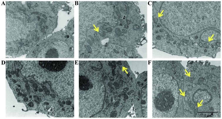
Figure 9.
Transmission electron microscopic observations (TEM) of LoVo WT and LoVo DX cells. (A-C) LoVo WT and (D-F) LoVo DX cells were incubated at 37°C for 60 min. (A and D) Untreated cells; (B and E) cells treated with 21 µM spermine alone; (C and F) cells treated with ZmPAO and 21 µM spermine. Scale bars, 1 µm. ZmPAO, maize polyamine oxidase; LoVo WT cells, LoVo wild-type cells; LoVo DX cells, LoVo multidrug-resistant cells.

