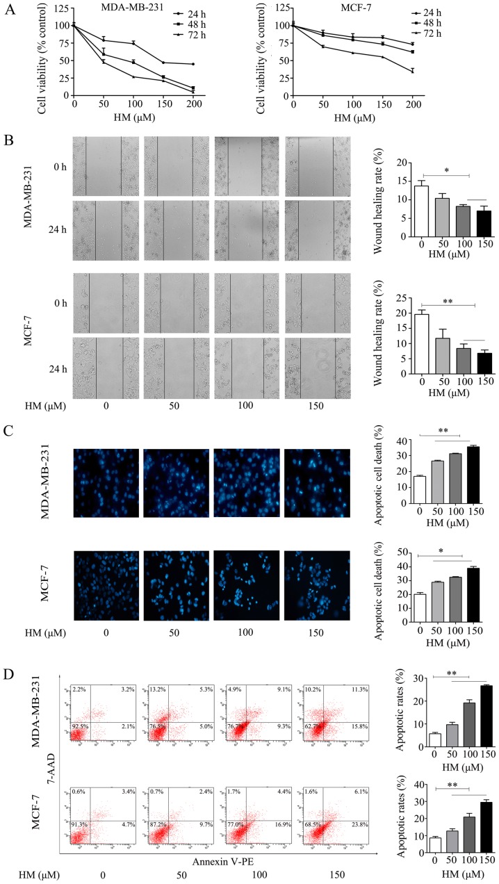
Figure 2.
HM inhibits proliferation and migration, and induces apoptosis of MDA-MB-231 and MCF-7 cells. (A) Dose- and time-dependent effects of HM on MDA-MB-231 and MCF-7 cell viability. Cells were treated with 50, 100 or 150 μM HM for 24, 48 and 72 h, and cell viability was measured via CCK-8 assay. Data are presented as the mean ± SD. (B) MDA-MB-231 and MCF-7 cells were treated with HM (50, 100 or 150 μM) for 24 h. Cell migration was detected using a monolayer wound healing assay (magnification, ×400). The wound healing rate at 24 h was quantified. (C) Apoptotic morphology of the MDA-MB-231 and MCF-7 cells was detected by fluorescent microscopy following DAPI staining (magnification, ×400). The histogram represents the apoptotic rate. (D) Analysis of apoptotic cells induced by HM for 24 h using flow cytometry. The histogram represents the apoptotic rates. Data are presented as the mean ± SD from three independent experiments. *P<0.05 and **P<0.01, with comparisons indicated by brackets. HM, harmine.

