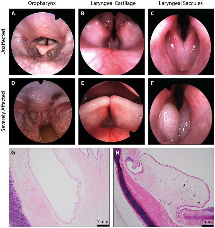
Fig 2. Pathological assessment of Norwich Terrier UAS.
(A-F) Images from the laryngoscopic videos that were used to grade anatomical components of the upper airway. Examples graded as ‘normal’ and ‘severely affected’ have been selected for each of the soft palate length (seen in the oropharynx), laryngeal cartilage position and laryngeal saccules. Histological preparations of the laryngeal ventricles from a (G) ‘normal’ and (H) ‘severely affected’ Norwich Terrier. Sections were made perpendicular to the ventricle, at the entrance to the larynx. Arrows indicate dilated lymphatic vessels.

