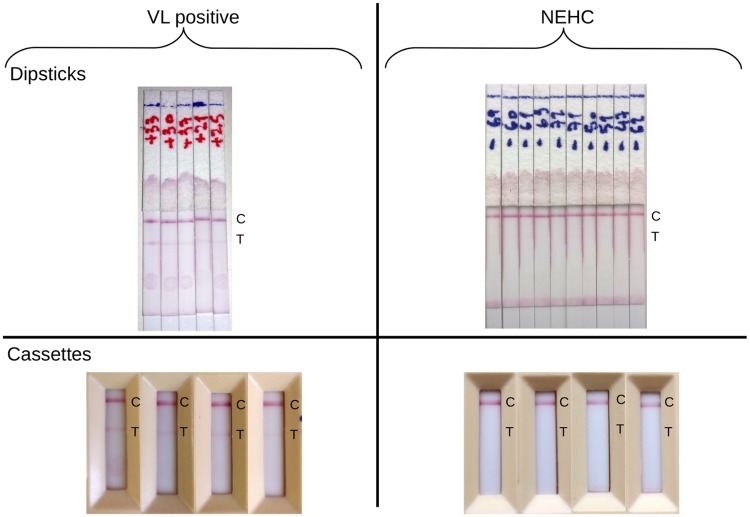
Fig 7. Peptide EpQ11 is specifically detected by Sudanese VL IgG1 in cassette as well as in dipstick format.
‘T’ indicates the location of the NLA at which, in a positive test, there is a red coloured line due to the presence of peptide-IgG1 complex. Successful migration was ensured by the development of the control line at ‘C’.

