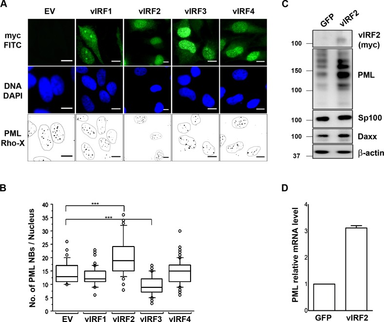
Fig 2. Effects of KSHV vIRF proteins on PML NBs.
(A) HeLa Cells were transfected with 1 μg of either the control vector or one of the vIRF expressing constructs and were fixed 36 h after transfection for IFA. Transfected cells express GFP, PML was detected with a mouse anti-PML antibody and a goat anti-mouse IgG Lissamine Rhodamine (LRSC)-conjugated secondary antibody and DNA was stained with DAPI. Images were acquired using a Carl Zeiss microscope at 100x magnification. For better visualization PML NBs are shown as black dots on a white background and the nuclei were encircled. Bars, 10 μm. (B) Quantification of immunofluorescence data from A, PML NBs were counted in at least 100 cells per each construct and a Man Whitney U test was performed to determine significance. Boxes indicate 25th to 75th percentile; central line inside each box indicates the median and the whisker illustrates the 5th and 95th percentile. The dots indicate the outliers. (C) HUVECs were transduced with either the control or the vIRF2 expressing lentiviral vector and 36 h after transduction cells were lysed and protein expression was analyzed by WB. (D) HUVECs were transduced with either the control or vIRF2 expressing lentivirus and 36 h later cells were lysed for RNA extraction. PML mRNA was quantified by reverse transcription following qPCR using dually labeled probes (Taqman).

