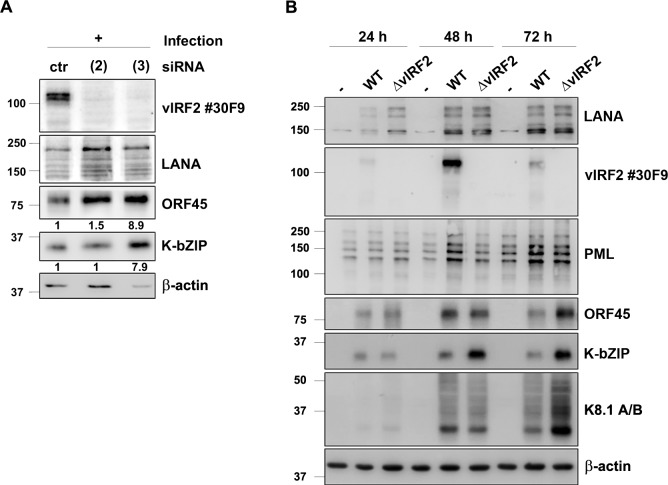
Fig 5. KSHV vIRF2 inhibits KSHV lytic protein expression after de novo infection.
(A) HuARLT cells were microporated with either a non-targeting siRNA (ctr) or with two different vIRF2 siRNAs (see Figs 3 and 4) and 24 h later cells were infected with rKSHV at a MOI of 5 (titer determined on HEK-293 cells). The cells were lysed 72 h after induction and protein expression was analyzed by WB. ORF45 and K-bZIP protein levels were quantified and normalized to β-actin levels by using the Image studio software. Relative expression levels (in comparison to those seen with the control siRNA) are indicated below the respective blots. (B) HuARLT cells were infected with either the KSHV.WT virus or the KSHV.ΔvIRF2 virus at a MOI of 5. The cells were lysed at the indicated time points and protein expression was analyzed by WB.

