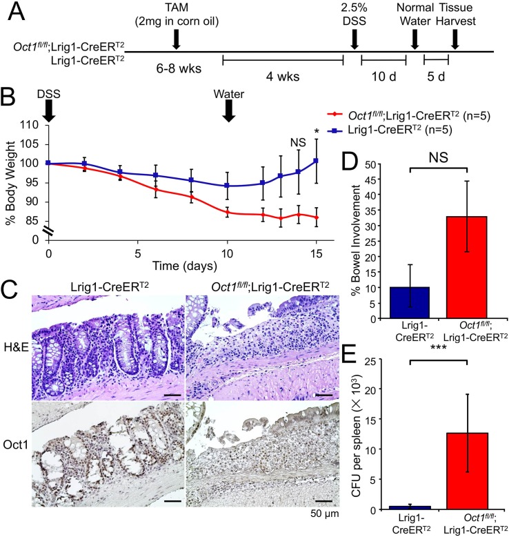
Fig 4. Loss of Oct1 renders the mouse colon more susceptible to DSS-induced damage.
(A) Experimental schematic. Lrig1-CreERT2;Oct1fl/fl mice and Lrig1-CreERT2 controls were treated with tamoxifen. 4 weeks post-tamoxifen treatment, mice were treated with 2.5% DSS in their drinking water for 10 days. All mice were female littermates. (B) Average body weight changes of the mice (n = 5 for each group from a single representative experiment) were monitored following DSS treatment. Error bars denote ±SEM. (C) Representative Oct1 IHC images of distal colon sections from mice following 10 day treatment with DSS and 5 day treatment with water. Above shows H&E stained adjacent sections of the same tissue. (D) Quantification across the entire colon (excluding cecum) of 5 mice in each group were evaluated for colitis involvement. Average involvement is shown. (E) Spleen homogenates from the same mice as in (D) were grown on blood agar plates to determine bacterial burden and loss of gut barrier function. Average CFUs are shown. N = 5 for each group.

