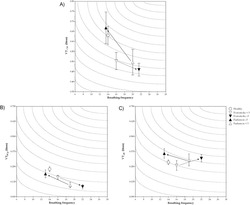
Fig 2. Minute ventilation of chest wall and rib cage pulmonary and abdomen compartments at rest in healthy individuals, post-stroke and Parkinson’s disease subjects according to length of disease diagnosis.
Data presented as median and interquartile range between 25–75%. A) minute ventilation of chest wall; B) minute ventilation of rib cage pulmonary compartment; C) minute ventilation of abdomen compartment; VTCW: tidal volume of chest wall (in liters); VTRCp: tidal volume of rib cage pulmonary compartment (in liters); VTAB: tidal volume of abdomen compartment (in liters).

