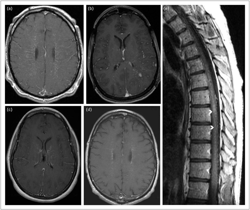
FIGURE 2.

Characteristic T1 postgadolinium MR images of autoimmune GFAP astrocytopathy (axial brain, a–d; sagittal spine, e). Patterns of brain enhancement include: (a) radial periventricular; (b) leptomeningeal and punctate; (c) serpiginous; and (d), periependymal. Spinal cord enhancement, e, is characteristically central, often adjacent to the canal (arrow heads). GFAP, glial fibrillary acidic protein; MR, magnetic resonance.
