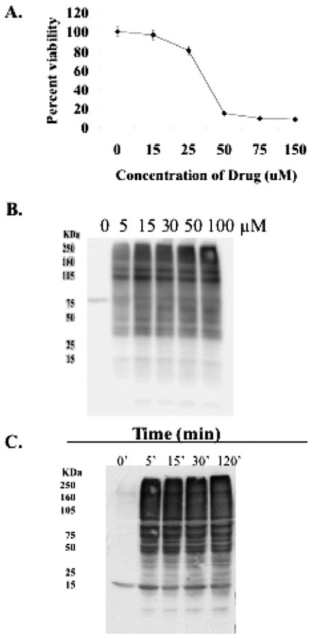
Fig. 3.
PABA/NO induces S-glutathionylation in a dose- and time-dependent manner. A, HL60 cells were treated with varying concentrations of PABA/NO. Cell viability was measured at 72 h with the MTT assay. B,1.5 × 106 HL60 cells were treated with varying concentrations of PABA/NO for 1 h. C, 1.5 × 10⁶ HL60 cells were treated with 30 μM PABA/NO for 0 to 120 min. After treatment, 20 μg of lysate was separated by SDS-PAGE and analyzed for S-glutathionylation by immuno-blot.

