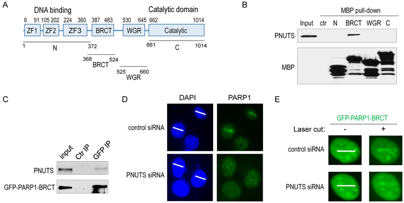
Figure 5. PNUTS knockdown impairs the recruitment of PARP1 to laser-induced DNA damage sites.
(A) The schematic diagram of PARP1 motifs and mutants. (B) As described in Materials and Methods, four segments of PARP1 (N: aa 1-372; BRCT: aa 368-524; WGR: aa 525-660; and C: aa 661-1014) were tagged with MBP, expressed in BL21 cells, and purified. The recombinant proteins were incubated in HeLa cell lysates. 20% input, control pull-down, and PARP1 pull-down samples were analyzed by immunoblotting for PNUTS and MBP. (C) As described in Materials and Methods, the BRCT domain of PARP1 was tagged with GFP and expressed in HeLa cells. GFP IP was performed. 20% input, control IP, and GFP IP samples were analyzed by immunoblotting for PNUTS and GFP. (D) HeLa cells with control or PNUTS siRNA (#1) treatment were microirradiated with 405nm laser. The path of laser microirradiation is marked by white lines (panels on the left). The localization of PARP1 3 min after laser treatment is shown by immunofluorescence (panels on the right). More than 10 cells were examined. (E) The BRCT domain of PARP1, tagged with GFP, was expressed in HeLa cells with control or PNUTS knockdown (#1). Cells were microirradiated with 405nm laser, and examined for the localization of GFP 3 min after laser treatment. More than 20 cells were examined.

