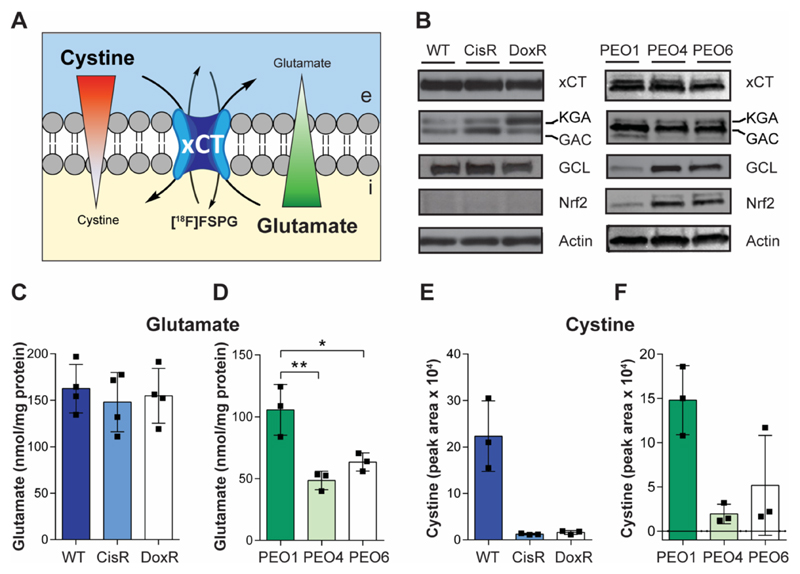
Fig. 3. Intracellular levels of cystine are decreased in drug-resistant cancer cells.
(A) Model of [18F]FSPG accumulation, with mechanisms known to control radiotracer accumulation. Uptake of [18F]FSPG is predicted to occur through exchange with intracellular glutamate, with efflux controlled by the exchange with extracellular cystine. Red and green triangles represent the concentration gradients of cystine and glutamate across the plasma membrane. e, extracellular; i, intracellular. (B) Western blot analysis of the levels of xCT, glutaminase isozymes (kidney-type glutaminase (KGA) and glutaminase-C (GAC)), glutamate-cysteine ligase (GCL) and NRF2. Intracellular levels of glutamate (C and D) and cystine (E and F) in A2780 (C and E) and PEO human ovarian cancer cell lines (D and F). Data presented as mean with individual scatter plots representing independent experiments. *, P < 0.05; **, P < 0.01.

