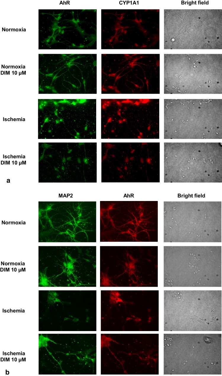
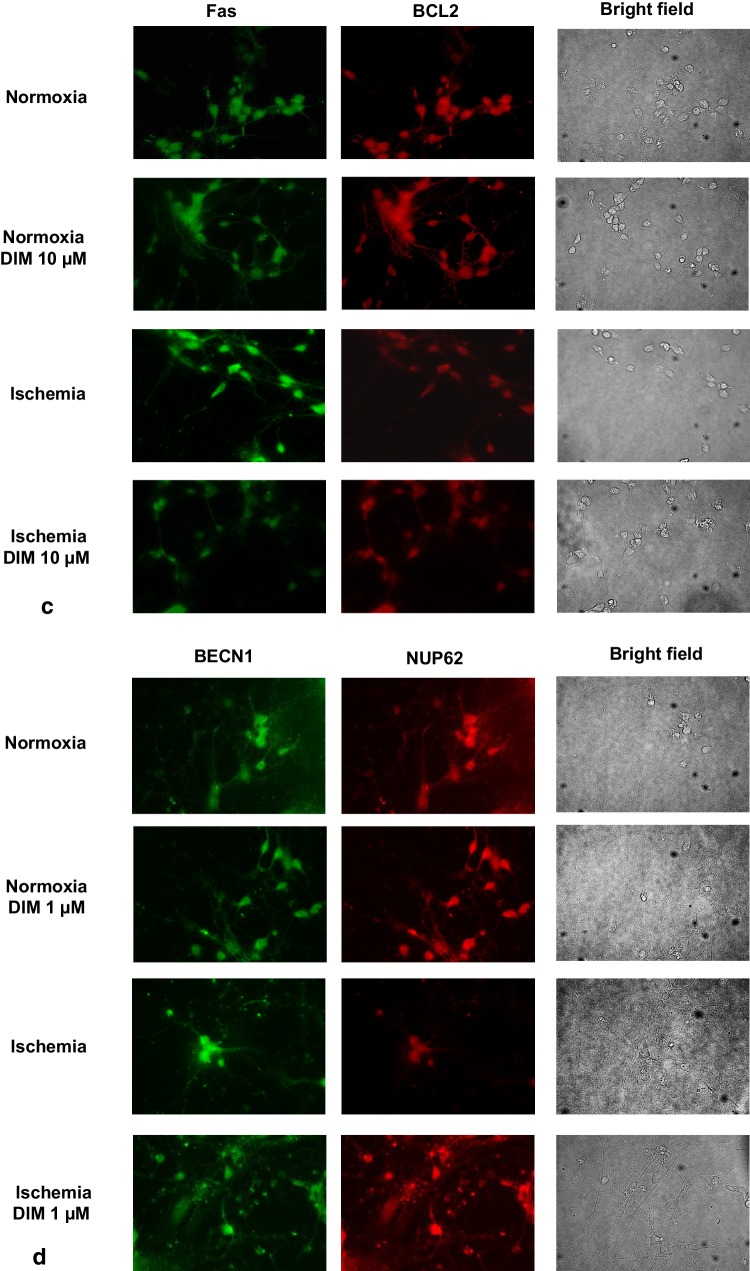
Fig. 14.
Impact of ischemia (6 h) and DIM (1,10 µM) on the cellular distribution of AhR (panel a—green, panel b—red), CYP1A1 (panel a—red), BECN1 (panel d—green) NUP62 (panel d—red), Fas (panel c—green), BCL2 (panel c—red) and MAP2 (panel b—green) in hippocampal cultures. Primary hippocampal cultures were treated with either ischemia alone or in combination with DIM for 6 h. The cells were cultured on glass cover slips and subjected to immunofluorescence double-labeling. The samples were analyzed using a confocal microscope (DMi8-CS, Leica MicroSystem, Wetzlar, Germany) with a HC Plan-Apochromat CS2 63x/1.4 Oil objective


