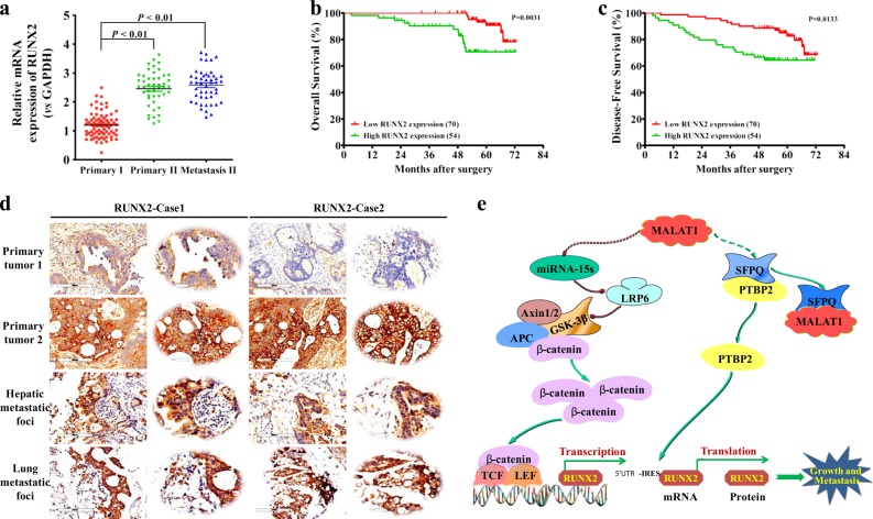
Fig. 8. RUNX2 overexpression correlates with CRC progression.
a Expression levels of RUNX2 in CRC tissues were analyzed by real-time PCR. The significant differences between primary tumor I (without paired metastatic tissues) and primary tumor II (with paired metastatic tissues) were analyzed using the Wilcoxon signed-rank test. **P < 0.01 (t test). b–c Kaplan–Meier analyses of the correlations between RUNX2 expression levels and OS or DFS of 124 CRC patients; the median expression levels were used as the cutoff. d Immunohistochemical analysis of RUNX2 proteins on consecutive tissue microarray slides of CRC tissues. Representative case 1 and case 2 were presented (scale bars, 200 and 50 mm, respectively). e A schematic model of MALAT1 regulating transcriptional and translational levels of RUNX2 in CRC metastasis

