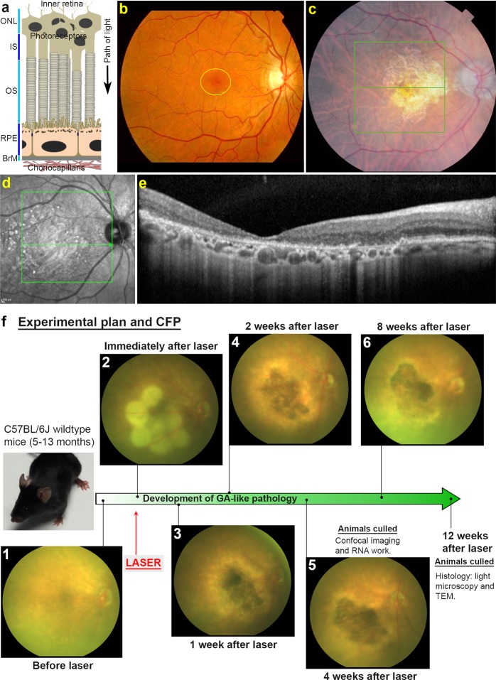
Figure 1.
The anatomy of geographic atrophy (GA) and development of GA-like pathology in lasered mice. (a) Schematic diagram of the outer retina showing arrangement of different cell layers. Outer nuclear layer (ONL), Photoreceptor IS (inner segments) and OS (outer segments), Retinal pigment epithelium (RPE), Bruch’s membrane (BrM) and Choriocapillaris. An arrow indicates the path of light. (b) A colour fundus photograph (CFP) of a healthy human retina with the macula encircled in yellow, (c) compared to a diseased retina from a GA patient. Notice presence of a central/macular lesion with defined borders. (d) Black and white optical coherence tomography scan of a GA lesion taken from a Heidelberg OCT, and (e) a cross-section (from central green line in d) showing respective layers of the outer retina affected by disease. (f) Development of GA-like pathology in lasered mice. Representative CFP from mice before and after laser and in subsequent weeks. Notice how multiple lasered spots in the central murine retina coalesce after 1 week to create a focused atrophic region. Longitudinal CFP imaging of a five month old mouse shows this GA-like lesion to persist for up to 8 weeks after laser treatment. The timeline of developing GA-like pathology alongside experimental end-points are also shown.

