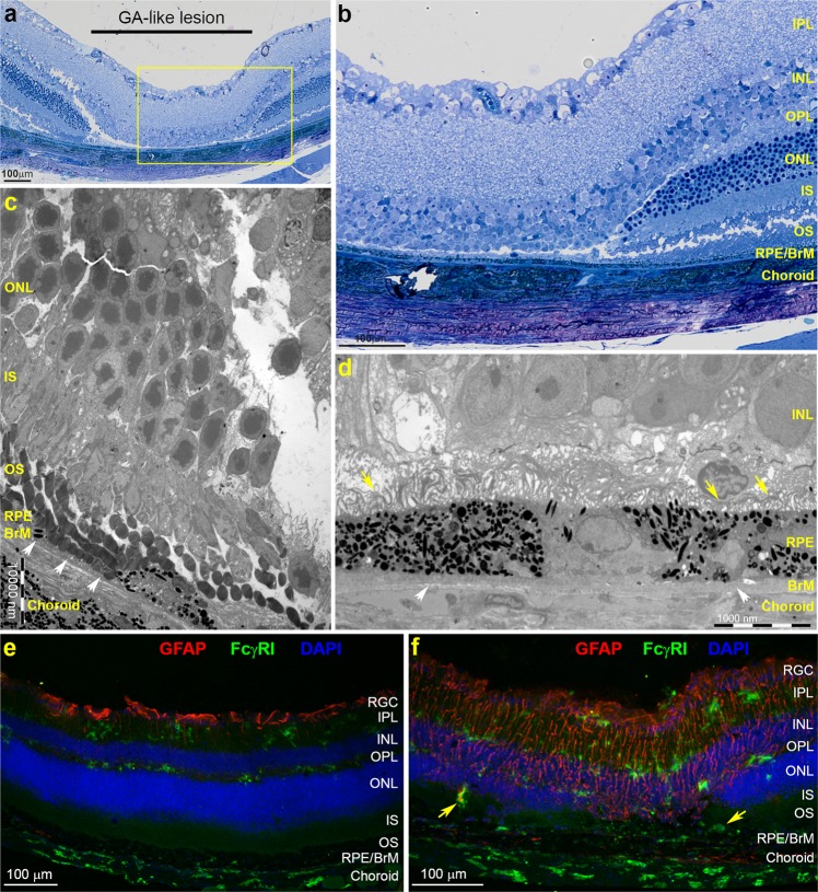
Figure 3.
Structural changes and chronic inflammation within lesions and adjacent tissues in lasered mice correspond to early GA-like pathology. (a) Representative semi-thin tissue sections from an eleven month old mouse at 3 months post-treatment and stained with Toluidine blue show a characteristic GA-like lesion where photoreceptors are absent in lasered areas. (b) Magnified insert from (a) (yellow box) where the INL is observed lying in close apposition to the RPE. The inner retina had collapsed into the area that had been occupied by the degenerated photoreceptor layer. Lesion margins could be observed as well-defined wedge-shapes, with photoreceptor IS/OS and ONL as well as the OPL taking on a more normal appearance further away from the lesion. The RPE and BrM remains intact alongside a choroid that also appears to be unaffected. Scale bars in A and B corresponds to 100 μm. (c) Representative electron micrograph showing pathology in lesion margins. Areas where RPE cells had become atrophic can be observed (white arrows) alongside photoreceptor OS horizontally positioned next to the underlying BrM. However, the RPE monolayer was intact in most micrographs. Importantly, the BrM itself appears to remain unaffected. Photoreceptor OS followed by IS and the ONL gradually tapers closer to the lesion. (d) Micrograph from within the lesion showing the INL next to the RPE in the absence of a photoreceptor layer. The RPE monolayer shows neighbouring hypopigmented as well as hyperpigmented cells. Microvilli on the apical RPE surface appear to be disorganised and shorter in length (yellow arrows), whilst the BrM shows varying thicknesses (white arrows). Scale bars in c and d correspond to 1000 nm. (e) Representative confocal-immunofluorescence comparing GFAP (red) and FcγRI (green) staining in non-lasered tissues vs. (f) adjacent lasered areas at 4 weeks post treatment. Notice GFAP staining in processes extending into what appears to be remaining photoreceptors within lesions after 4 weeks, which had disappeared by 12 weeks when visualised by light and electron microscopy. GFAP expression was also observed in the INL and IPL layers, extending beyond the lesion into surrounding tissues. No GFAP expression was observed in non-lasered tissues except for constitutive staining near the vitreous interface. Upregulated FcγRI expression was observed clustered within lesions and in marginal tissues (yellow arrows). Only minimal FcγRI expression was observed in non-lasered tissues. Nuclei were labelled with DAPI and appear blue. Scale bars in e and f correspond to 100 μm. Retinal ganglion cells (RGC), Inner plexiform layer (IPL), Inner nuclear layer (INL), Outer plexiform layer (OPL), Outer nuclear layer (ONL), IS/OS (inner and outer segments) of photoreceptors and Retinal pigment epithelium (RPE).

