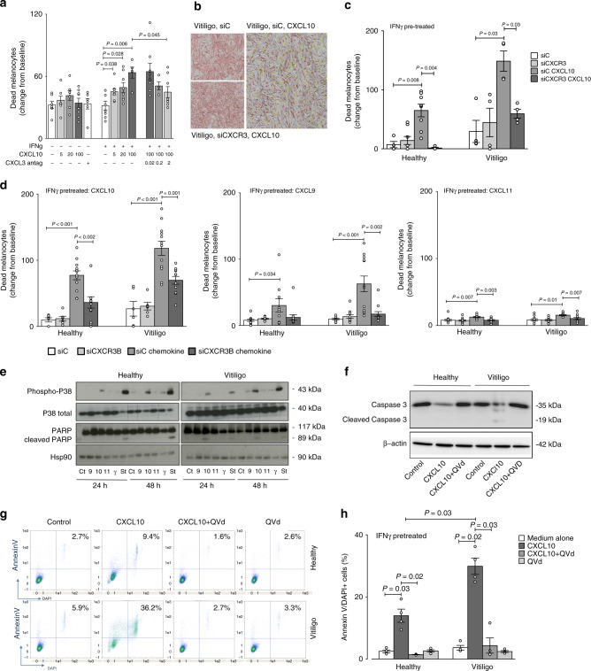
Fig. 4.
CXCL10 activates melanocytic CXCR3B to induce apoptosis. a Effect of CXCL10 (5, 20, 100 pg/ml) on cell viability in unstimulated or IFNγ stimulated (50 ng/ml for 48 h) melanocytes from vitiligo patients, in presence or absence of CXCR3 antagonist AS612568 (0.02 μM, 0.2 μM or 2 μM) (n = 5–8). Cell viability was monitored using lncuCyte® live cell fluorescence imaging system. b Illustrates live IncuCyte images of vitiligo melanocytes 24 h after exposure to CXCL10 in cells transfected with siC or siCXCR3. Melanocytes were tracked with CellTrackerTM Red CMPTX dye and dead cells tracked with IncuCyte® Cytotox Green reagent. Co-localised yellow cells represent dead melanocytes. The effect of siCXCR3 (or its siC) on CXCL10 (100 pg/ml)-induced death of healthy (n = 4–8) and vitiligo (n = 4) melanocytes are shown in c. In d healthy and vitiligo melanocytes (n = 6–12) were transfected with siCXCR3B (or its siC) and melanocyte death shown at 24 h following CXCL10, CXCL9 or CXCL11 (100 pg/ml) stimulation. In separate experiments, cell lysates was used to study the signalling pathway induced by chemokines, IFNγ (50 ng/ml) or Staurosporine (positive control, 1 μg/ml) at 24 or 48 h post stimulation, measuring the expression levels of phosphorylated and total p38 and total and cleaved poly(ADP-ribose) polymerase (PARP) by Western Blot analysis (e). HSP90 was used as an internal loading control. Representative blot of 3 separate experiments is shown. Total and cleaved caspase-3 activity (f) and proportion of apoptotic cells (counted as % Annexin V+DAPI+cells in FACS analysis) (g) from healthy (n = 4) and vitiligo (n = 4) melanocytes stimulated with 100 pg/ml CXCL10 for 24 h in presence or absence of QVd OPh (10 μM, caspase inhibitor) (h). Results are shown as individual dot plots with a line at mean ± SEM

