Fig. 5.
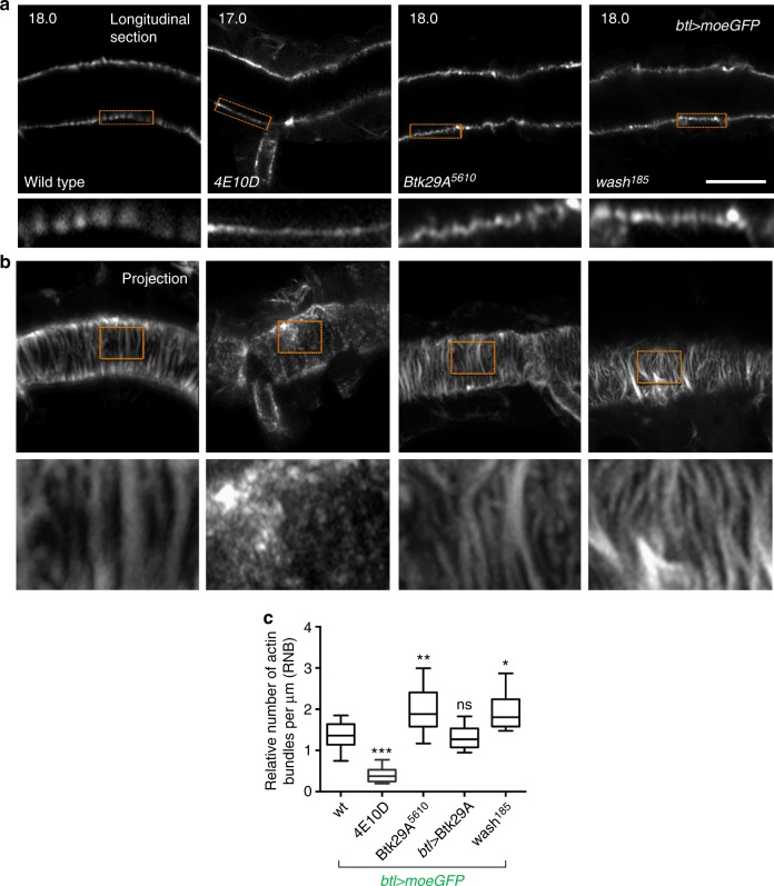
A new role of protein tyrosine phosphatases (PTPs) in actin cytoskeleton regulation. a, b Airyscan confocal images of the dorsal trunk of wild-type, Ptp4EPtp10D, Btk29A5610 and wash185 embryos expressing the actin reporter btl>moeGFP (grey). Images were acquired during luminal endocytosis, 18 h after egg laying (AEL) (for wild-type, Btk29A5610 and wash185) and 17 h AEL (for Ptp4EPtp10D). Longitudinal sections (a) and Z-stack projections (b) are shown. Lower rows in a and b depict zoomed view of areas indicated by the rectangular frames. Scale bars, 10 μm. c Plots showing the relative number of actin bundles (>2 μm long) per μm (RNB) in wild type (n = 10), Ptp4EPtp10D (n = 8), Btk29A5610 (n = 14), btl>Btk29A (type-2) (n = 11) and wash185 (n = 14) embryos expressing btl>moeGFP. The boxplot shows the median (horizontal line) and the data range from 25th to 75th percentile. The bars denote maxima and minima values. *P < 0.01, **P < 0.001, ***P < 0.0001 indicates statistical significance in comparison to wild-type (unpaired two tailed t tests)

