Fig. 8.
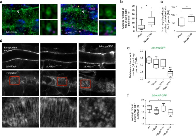
The WASHY273 regulates actin organization and luminal clearance. a Airyscan confocal sections showing the dorsal trunk (DT) of btl>Wash or btl>WashY273D embryos expressing the actin cytoskeleton reporter moe-GFP stained with anti-GFP (green), anti-Rab7 (magenta) and DAPI (blue). Images on the right of each confocal section depict zoomed views of Rab7-positive ring-shaped actin patches (arrowheads). b, c Plots showing the average number of ring-shaped actin patches in btl>Wash (n = 7) or btl>WashY273D (n = 13) at 18 h after egg laying (AEL) (b), and the percentage of ring-shaped actin patches in btl>Wash (n = 8) or btl>WashY273D (n = 14) colocalized with Rab7 puncta at 18 h AEL (c). Error bars show s.e.m. *P < 0.02 (unpaired two tailed t tests). d Airyscan confocal sections showing the DT of wild-type living embryos expressing btl>Wash or btl>WashY273F or btl>WashY273D and moe-GFP (grey) (18 h AEL). Images of the lowest row are zoomed areas of the cortical cytoskeleton indicated by the red rectangular frames. e Plots showing the relative number of actin bundles (>2 μm long) per μm (RNB) in wild-type (n = 14) and embryos expressing btl>Wash (n = 18) or btl>WashY273F (n = 15) or btl>WashY273D labelled by btl>moeGFP (n = 22). **P < 0.002 (unpaired two tailed t tests). f Plots showing average time (hours) of the luminal ANF-GFP clearance in wild-type (n = 66), btl>Wash (n = 38) or btl>WashY273F (n = 39) or btl>WashY273D (n = 41) embryos. **P < 0.001 compared to wild type (unpaired two tailed t tests). The boxplots (b, c, e, f) show the median (horizontal line) and the range from 25th to 75th percentile. The bars depict maximum and minimum values. Data collected from 5–7 independent experiments

