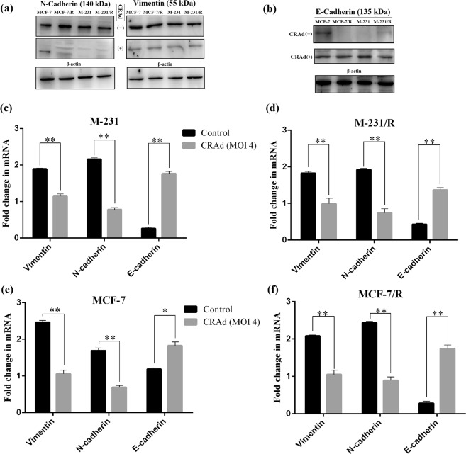
Figure 7.
CRAd regulates cancer cell migration through EMT reversal: Breast cancer cells (MCF-7 and M-231) and their resistant sublines (MCF-7/R and M-231/R) were treated with CRAd for 48 hr at an MOI of 5 and subsequently lysed and treated for protein expression analysis via blotting. (a,b) Western blot analysis shows that CRAd treatment (+) represses mesenchymal markers, vimentin and N-cadherin, while restores epithelial marker, E-cadherin expression. Controls (un-treated cells) showed opposite trends. The data shown above are the average of triplicate experiments (p < 0.05) and blot images were cropped for clarity of the presentation. (c–f) qRT-PCR detection of mRNA levels of EMT markers in human breast cancer cell lines upon CRAd treatment. The grouped blots were cropped from different parts of the same gel. Uncropped blots are shown in the Supplementary Information (See Supplementary Data, Figs S1 and S2).

