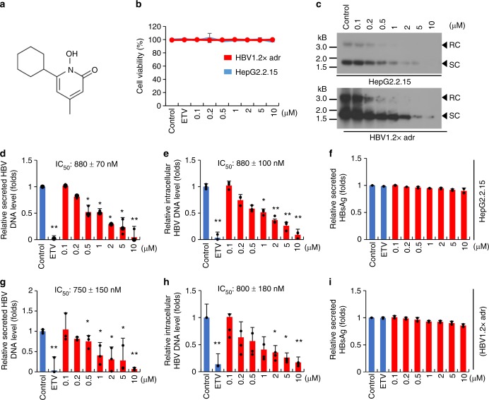
Fig. 4.
Ciclopirox inhibits HBV replication. a Chemical structure of ciclopirox. b HepG2.2.15 cells (blue), and HepG2 cells transfected with pHBV1.2× (red) were treated with various concentrations (0.1–10 μM) of ciclopirox for 6 days and assayed for cytotoxicity. c Intracellular HBV DNA was detected by southern blot analysis. d–i Secreted HBV DNA (d, g, respectively) and intracellular HBV DNA (e, h, respectively) were measured by quantitative PCR. Secreted HBsAg (f, i respectively) was quantified by ELISA. The data in b–i are representative of two or three independent experiments and are expressed as mean ± SD. The error bars represent the ± SD. *p < 0.05; **p < 0.01, by unpaired two-tailed Student’s t-tests. Source data are provided as a Source Data file

