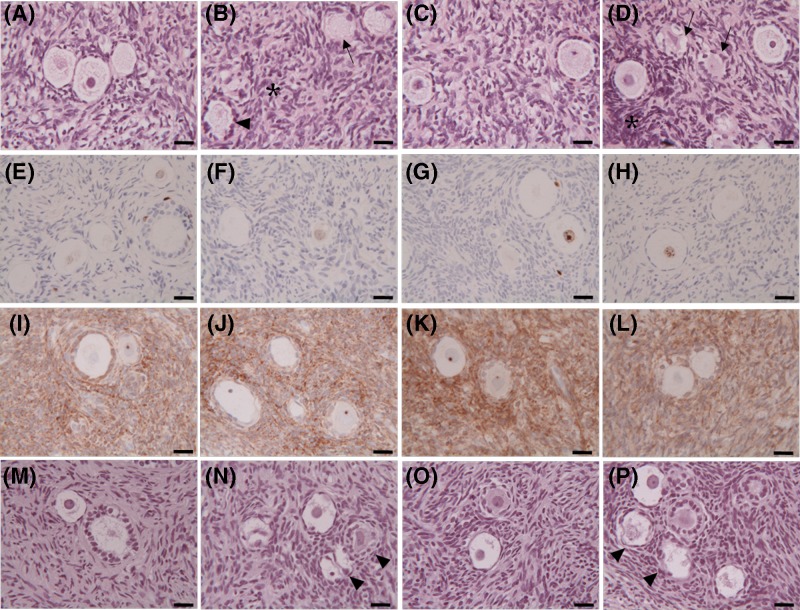
Figure 7. Effects of DOX and EGCG on preservation and viability of the ovarian tissue.
Light microscopy of ovarian tissue after 24 h treatment with DOX 1 μg/ml, EGCG 10 μg/ml, and combined treatment with DOX+EGCG at the aforementioned concentrations. (A–D) Hematoxylin/eosin staining of untreated tissue-CTR (A) and treated with DOX (B), with EGCG (C), with DOX+EGCG (D). (E–H) Immunohistochemical staining for ki-67 of CTR tissue (E) and tissue treated with DOX (F), with EGCG (G), with DOX+EGCG (H). (I–L) Immunohistochemical staining for Bcl2 of CTR tissue (I) and tissue treated with DOX (J), with EGCG (K), with DOX+EGCG (L). (M–P) TUNEL assay of untreated tissue-CTR (M) and treated with DOX (N), with EGCG (O), with DOX+EGCG (P). Irregular-shaped follicles (arrowheads); follicles with cytoplasmic vacuoles (arrows); areas with pyknotic nuclei of stromal cells (asterisks). Scale bar = 25 µm.

