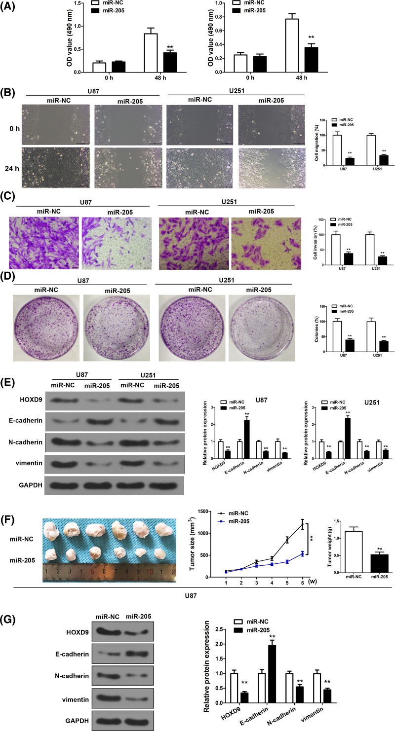
Figure 2. miR-205 overexpression inhibits invasive ability of U251 and U87 cells and regulates EMT-related gene expression.
(A) Overexpression of miR-205 decreased U87 and U251 cell growth. (B) Wound-healing assay in U87 and U251 cells overexpressing miR-205. The wound gaps were photographed and measured. (C) Transwell invasion assay of U87 and U251 cells overexpressing miR-200b. Cells in the bottom of the invasion chamber were fixed, stained, and photographed. (D) Colony formation assays after 2 weeks of miR-205 treatment. (E) Western blot analysis was conducted to examine the protein expression of HOXD9, N-cadherin, E-cadherin, and Vimentin from U87 and U251 cells. (F,G) MiR-205- or miR-NC- treated U87 cells were implanted subcutaneously into nude mice and tumor formation was measured. (F) Tumor volume was measured once a week. And tumor weight was determined at 6 weeks after cell injection (n=6) (G) Western blot analysis was conducted to examine the protein expression of HOXD9, N-cadherin, E-cadherin, and Vimentin from the tumor tissues. Assays were performed in triplicate. **P< 0.01, means ± S.D. was shown.

