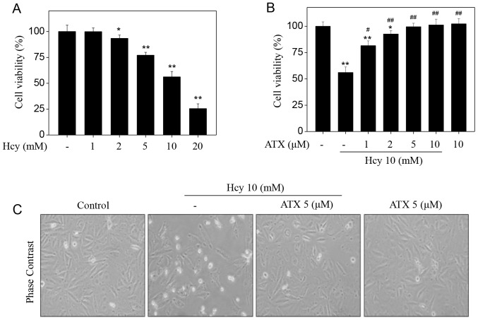
Figure 1.
ATX inhibits Hcy-induced cytotoxicity in HUVECs. (A) Cytotoxicity of Hcy towards HUVECs. Cells (8,000 cells/well) were seeded in 96-well plate and treated with Hcy for 72 h. (B) ATX pre-treatment inhibited Hcy-induced HUVEC cytotoxicity. Cells were pretreated with 1–10 µM ATX for 6 h and co-treated with 10 mM Hcy for 72 h. Cell viability was detected by an MTT assay. (C) Morphological changes of HUVECs. Following treatment, cells were observed under a phase-contrast microscope (magnification, ×400). All data and images were obtained from three independent experiments. *P<0.05, **P<0.01 vs. control; #P<0.05, ##P<0.01 vs. Hcy-treated group. ATX, astaxanthin; Hcy, homocysteine; HUVECs, human umbilical vascular endothelial cells.

