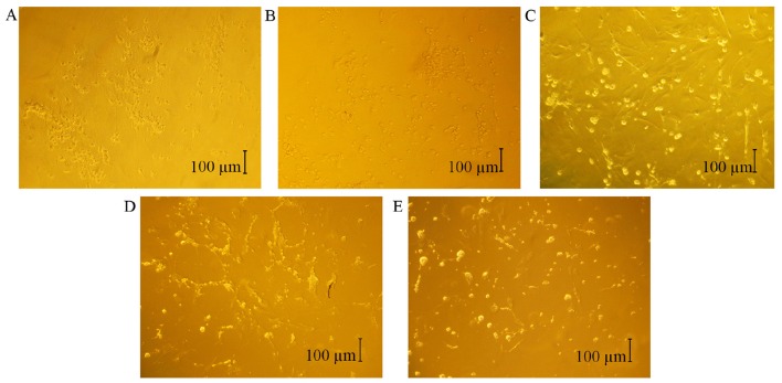
Figure 2.
Elevated HP induced apoptotic morphological changes in RGCs in vitro. The effect was observed by inverted phase contrast microscopy. (A) Micrograph of RGCs cultured at HP values of 0 mmHg (magnification, ×100). (B) Micrograph of RGCs cultured at HP values of 20 mmHg (magnification, ×100). (C) Micrograph of RGCs cultured at HP values of 40 mmHg (magnification, ×100). (D) Micrograph of RGCs cultured at HP values of 60 mmHg (magnification, ×100). (E) Micrograph of RGCs cultured at HP values of 80 mmHg (magnification, ×100). HP, hydrostatic pressure; RGC, retinal ganglion cell.

