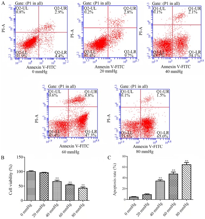
Figure 3.
Elevated HP reduced viability and induced apoptosis in RGCs in vitro. These effects were observed using a Cell Counting K-8 assay and by Annexin V FITC/PI flow cytometry, respectively. (A) Annexin V FITC/PI flow cytometry results at different HP values (0, 20, 40, 60 and 80 mmHg). (B) Quantification of cell viability rates. (C) Quantification of cell apoptosis rates. Data are presented as the mean ± standard deviation (n=3). **P<0.01 vs. the 0 mmHg group. HP, hydrostatic pressure; RGC, retinal ganglion cell; PI, propidium iodide; FITC, fluorescein isothiocyanate; UL, upper left; UR, upper right; LL, lower left; LR, lower right.

