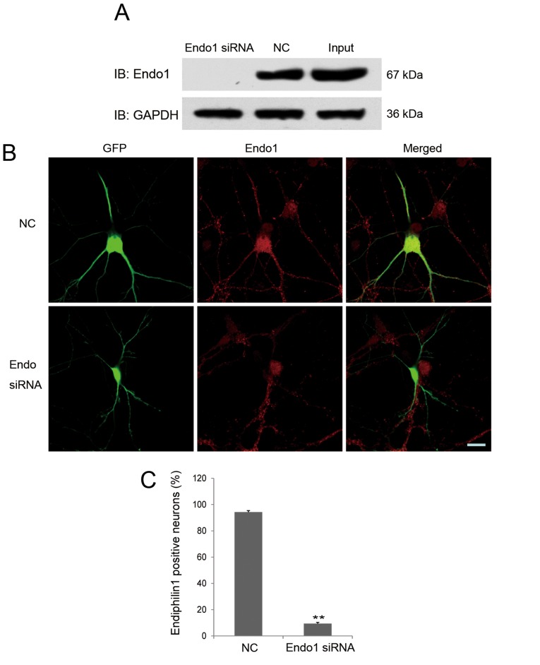
Figure 3.
Verification of the efficacy of endophilin 1 siRNA. (A) 293 cells were co-transfected with endophilin 1-pEGFP-C1 plasmids and Endo1 siRNA or NC. After 48 h, endophilin 1 and GAPDH as a control in the cell lysates were probed by the designated antibodies. (B) Images of neurons co-transfected with green fluorescent protein and NC or Endo1 siRNA. Cultures were fixed after transfection for 2–3 days and stained using endophilin 1 antibody (red). Scale bar, 20 µm. (C) Percentage of endophilin 1-positive neurons in the two groups. n=4, **P<0.01. Endo1, endophilin 1; siRNA, small interfering RNA; NC, negative control; IB, immunoblot; GFP, green fluorescent protein; Input, endophilin 1-pEGFP-C1 plasmid transfection only.

