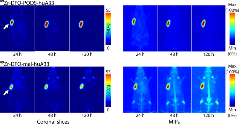
Figure 3.
Planar (left) and maximum intensity projection (right) PET images of athymic nude mice bearing A33 antigen-expressing SW1222 colorectal cancer xenografts (white arrow) following the injection of 89Zr-DFO-PODS-huA33 and 89Zr-DFO-mal-huA33 (140 μCi, 60–65 μg). The coronal slices intersect the center of the tumors.

