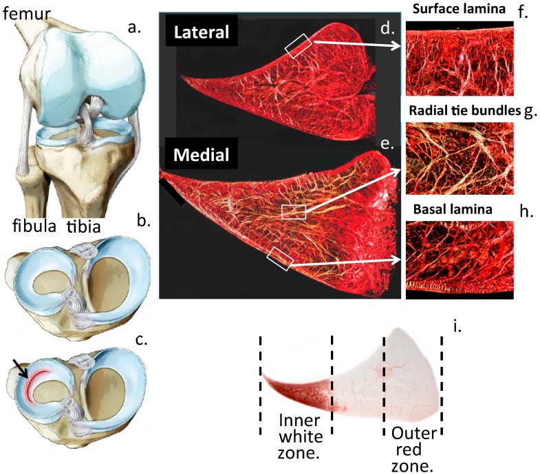
Figure 1.
Knee anatomy showing the location of the knee joint menisci (a, b) with a radial tear arrowed (c) and the collagen fibre organisation of the menisci in vertical sections through lateral (d) and medial menisci (e) which equip these as weight bearing stabilising structures in the knee joint. Radial tie bundles and collagen fibrillar arrangements in the surface and basal meniscal laminas are also shown (f–h). The inner zone meniscal cells are cartilaginous whereas the outer zone meniscal cells are more fibrocartilaginous (i). This is clearly evident in the focal localisation of the HS-proteoglycan perlecan which is a chondrogenic marker [10]. Figure modified from [18] with permission of InTech Open Publishers: London. Figure copyright J. Melrose.

