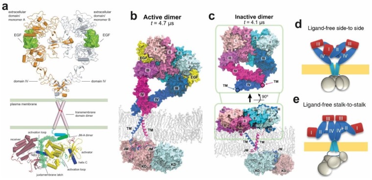
Figure 6.
(a) Full EGFR model in which the ECM and intracellular module (ICM) meet across the membrane, the only gap being the short outer JM segment. Taken, with permission, from Jura et al. [177]. (b,c) simulation of the near full-length active and inactive dimers, respectively. Taken, with permission, from Arkhipov et al. [67]. (d,e) models for alternative dimers produced by a combination of high-resolution imaging methods, including fluorescence localization imaging with photobleaching (FLImP), Förster resonance energy transfer (FRET), and single particle tracking and atomistic MD simulations. Taken, with permission, from Zanetti-Domingues et al. [50].

