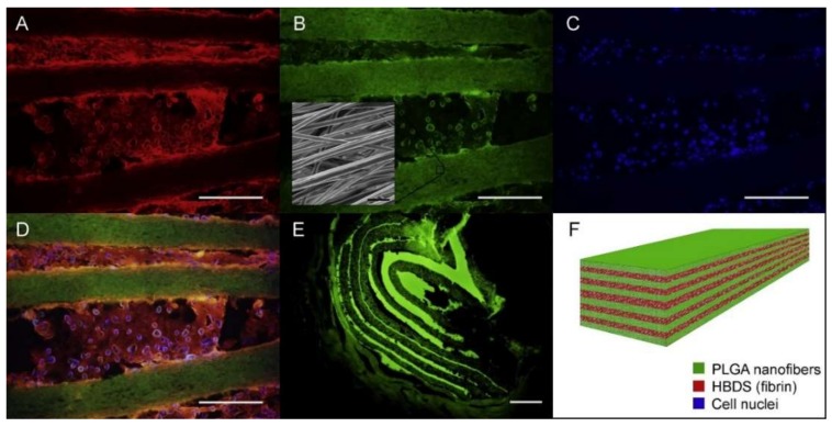
Figure 7.
A representative HBDS/nanofiber scaffold with 11 alternating layers of aligned electrospun PLGA nanofiber mats separated by HBDS containing 1 × 10⁶ ASCs is shown. (A–D) Micrograph showing the HBDS/nanofiber scaffold in vitro; the PLGA is labeled with FITC (green), the HBDS is labeled with Alexa Fluor 546 (red), and the ASC nuclei are labeled with Hoescht 33258 (blue) (scale bar = 200 μm). (B, inset) SEM image of the scaffold showing PLGA nanofiber alignment. (E) Micrograph showing the HBDS/nanofiber scaffold in vivo nine days after implantation in tendon repair. Eleven alternating layers of PLGA and HBDS can be seen (i.e., six layers of PLGA and five layers of fibrin); the PLGA is labeled with FITC (green) (scale bar = 100 μm). (F) A schematic of the layered scaffold is shown [86]. Reproduced with permission from Elsevier, 2013.

