Figure 2.
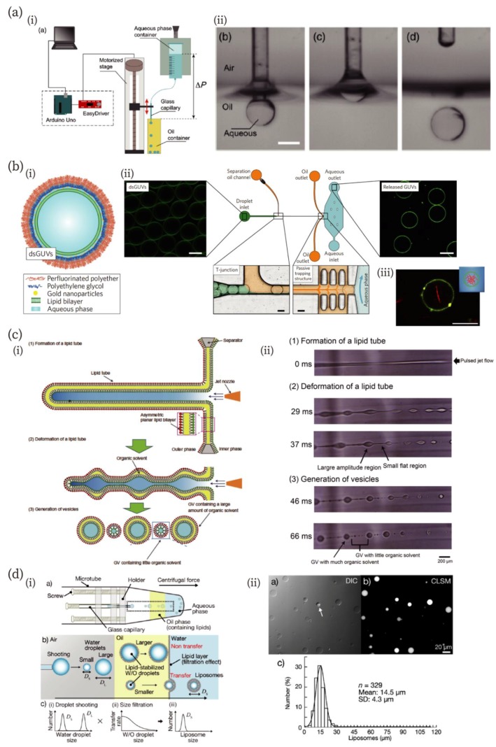
Novel and unique microfluidic technologies improving the fabrication of artificial cell-compartments. (a) A low-cost capillary-based fabrication method of W/O emulsion [66]. (i) An illustration of the low-cost setup. (ii) Snapshots of processes of droplet fabrication. Scale bar: 400 µm. Reproduced with permission from [66]. (b) A microfluidic chip for the fabrication of droplet-stabilized giant unilamellar vesicles (dsGUVs) and for the release of GUVs from the droplet [67]. (i) A schematic of the dsGUV. (ii) The microfluidic device used to release the GUVs from the surrounding stabilizing polymers into an aqueous phase. (iii) A fluorescence microscopic image of GUVs encapsulating actin filaments (red). Scale bars: 20 µm. Reproduced with permission from [67]. (c) A jetting-based fabrication method for asymmetric GUVs containing little organic solvent in the membrane [71]. (i) and (ii) Schematics and microscopic images of the formation process, reproduced with permission from [71]. (d) Droplet-shooting and size-filtration (DSSF) method for the fabrication of the compartment [73]. (i) Schematics of the device and formation process of GUVs. (ii) Differential interference contrast (DIC) and confocal laser scanning microscopic (CLSM) images of GUVs, and size-distribution of GUVs. The white arrow in the DIC image indicates a multilamellar vesicle. Reproduced with permission from [73].

