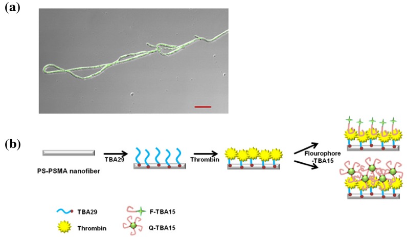
Figure 2.
(a) Composite microscope picture combining the brightfield and fluorescence of anti-thrombin aptamer sandwich on nanofiber with a quantum dot-labeled secondary aptamer (scale bar: 20 ); (b) schematic of the nanowire-based anti-thrombin sandwich assay (TBA: thrombin-binding aptamer). Reprinted from [20], Copyright 2012, with permission from Elsevier.

