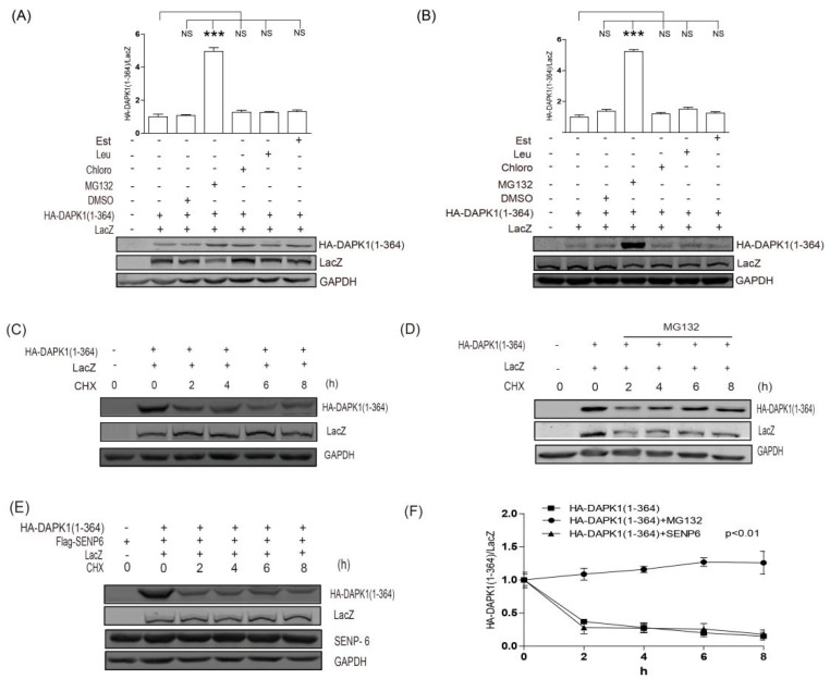
Figure 4.
SUMO pathway did not regulate the protein degradation of HA-DAPK1 (1–364). (A) HEK293T cells or (B) HCT116 cells transfected with DAPK1 (1–364) and LacZ were exposed to MG132 (10 μM, 6 h) or leupeptin (200 μM), Est (10 μg/mL) and chloroquine (100 μM) for 24 h as indicated. (C) HEK293T transfected with LacZ, DAPK1(1–364) were exposed to 20 μg/mL CHX for 0–8 h as indicated. (D) HEK293T transfected with HA-DAPK1 (1–364) and LacZ were exposed to 10 μM MG132 and 20 μg/mL CHX for 0–8 h as indicated. (E) HEK293T transfected with LacZ, HA-DAPK1 (1–364) and Flag-SENP6 were exposed to 20 μg/mL CHX for 0–8 h as indicated. The statistical results of (C–E) are summarized in (E). Cell lysates were extracted and immunoblotted with respective antibodies. The intensity of the bands was quantified using LICOR Odyssey software and represented by trend lines. The experiments were repeated three times (n = 3) and representative images are presented. The representative western blot images are from different gels and each lane was loaded with the same amount of proteins. Data of triplicate assays are presented as mean ± S.D. * p <0.05; ** p < 0.01; *** p < 0.001; NS, no significance. “+” indicated that the plasmid was transfected, “−” indicated that the plasmid was not transfected.

