Figure 3.
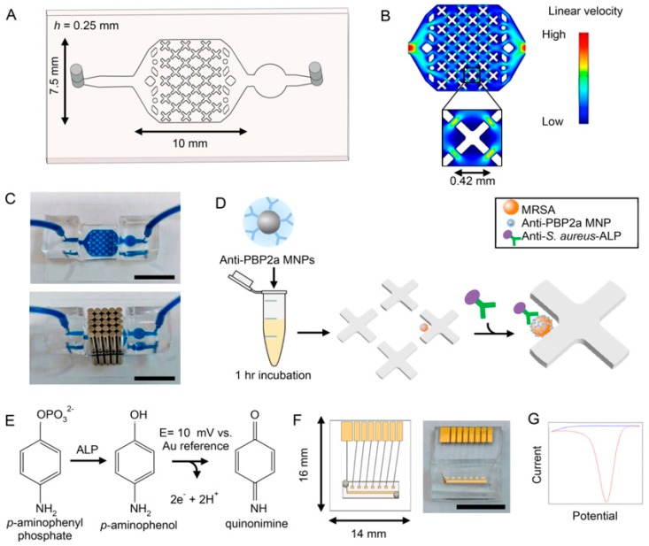
Bacterial capture and electrochemical detection. (A) Schematic of bacterial capture device fabricated in PDMS. (B) Flow profile of capture device simulated on COMSOL Multiphysics. X-shaped features create areas of reduced flow velocity. (C) Photograph of bacterial capture device filled with dye in the absence, and presence of, an array of external magnets (above and below images, respectively). Scale is 10 mm. (D) Filtered nasal swab specimen is incubated with anti-PBP2a MNPs for 1 h. The solution is then processed with the device, where magnetically-labeled bacteria are captured in areas of low flow velocity. After wash steps, anti-S. aureus antibodies functionalized with ALP are introduced into the device and washed. (E) The substrate p-APP is introduced to the device, where it is converted to electrochemically active p-AP by ALP. p-AP is oxidized to quinonimine at a potential of 10 mV against a gold reference electrode. (F) Schematic (left) and photograph (right) of electrochemical detector chip. A PDMS channel allows simple transfer of electrochemical readout solution from the capture device. Detection utilizes on-chip working and reference gold electrodes and an external Pt counter electrode. Scale is 10 mm. (G) Differential pulse voltammogram displaying signal from p-APP (blue) and p-AP (red). The measured current correlates to number of captured bacteria. Reprinted with permission from [83]. Copyright (2019) American Chemical Society.

