Figure 1.
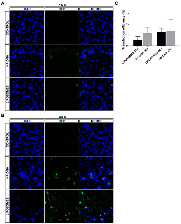
Analysis of transfection efficiency of DNA-wrapped gold nanoparticles (40 nm) compared to liposomes in differentiated ARPE-19 cells. Representative images of differentiated ARPE-19 cells transfected with the pEGFP reporter vector using either liposomes (LIPOTRANSFECTINE) or nanoparticles at (A) 16 h and (B) 48 h (for wider field images with a lower amplification, see Supplementary Materials, Figure S1). (C) Quantification of green fluorescent protein (GFP)-positive cells showed similar levels of transfection efficiency by nanoparticles (1.4% positive cells using 0.25 μg/150,000 cells) compared to standard lipofection (1.31% positive cells using 0.5 μg/150,000 cells) at 48 h. Remarkably, nanoparticles promoted GFP expression in transfected cells at an earlier time after transfection compared to liposomes, since at 16 h, 1.2% cells were GFP-positive in nanoparticle-transfected cells compared to 0.54% in those transfected with liposomes, thus suggesting different cellular uptake and/or intracellular vesicular trafficking routes for the two transfection systems. Quantification on 1100–1600 cells per condition.

