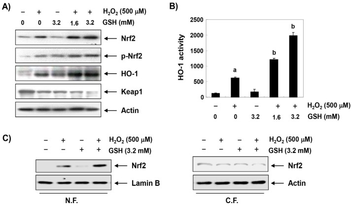
Figure 3.
Effects of glutathione on the expression of Nrf2 and HO-1 in H2O2-treated RAW 264.7 cells. RAW 264.7 cells were pretreated with the indicated concentrations of glutathione for 1 h and then stimulated with or without 500 μM H2O2 for 1 h. (A) Western blot analyses were performed with the indicated antibodies. The proteins were visualized using an enhanced chemiluminescence (ECL) detection system. Actin was used as an internal control. (B) The nuclear and cytosolic proteins were prepared and followed by Western blotting using the indicated antibodies. Lamin B and actin were used as internal controls. N.F., nuclear fraction; C.F., cytosolic fraction. (C) The HO-1 activities of cells grown under the same conditions were determined based on the bilirubin formation. The data were shown as the mean ± SD obtained from three independent experiments (a p < 0.05 compared with the control group; b p < 0.05 compared with the H2O2-treated group).

